Saphenous Nerve Entrapment Neuropathy
By Brad McKechnie, DC, DACAN
Saphenous nerve neuropathy is an important consideration in the differential diagnosis of lumbar radiculopathy, medial knee pain, and lower extremity vascular insufficiency. The saphenous nerve is the largest cutaneous branch of the femoral nerve and is purely sensory, with branches of the L3 and L4 nerve root levels contributing to the nerve.
The nerve is comprised of two functional divisions; the infrapatellar branch, and the descending branch.1
The saphenous nerve exits from the adductor canal (a.k.a. Hunter's canal, subsartorial canal), descends under the sartorius muscle, and then winds around the posterior edge of the sartorius muscle at its tendon portion. The infrapatellar branch pierces the sartorius muscle and courses anteriorly to the infrapatellar region. The descending branch passes down the medial aspect of the leg and, at the lower third of the leg, divides into two branches. One of the branches of the descending portion of the saphenous nerve courses along the medial border of the tibia and ends at the ankle while the other branch passes anterior to the ankle and is distributed to the medial aspect of the foot, sometimes reaching as far as the metatarsophalangeal joint of the great toe.1
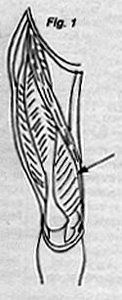
The adductor canal, the entrapment site for the saphenous nerve, is located approximately 10 centimeters (four finger widths) proximal to the medial femoral condyle. It is located by palpating along the anteromedial aspect of the vastus medialis muscle and then sliding posteriorly until the edge of the sartorius muscle is felt. The adductor canal is located directly beneath this point (See figure 1).2,4
Chief complaints associated with saphenous nerve entrapment include medial knee and/or leg pain after prolonged walking or standing and pain in the distribution of the saphenous nerve following quadriceps exercise.5Pain associated with this entrapment syndrome is characterized as burning by most patients and may be present at rest. In one study,2 two thirds of affected patients complained of pain at night. Additionally, stair climbing may aggravate symptoms associated with this entrapment neuropathy.
Clinical criteria for the diagnosis of saphenous nerve entrapment neuropathy includes pain in the distribution of the saphenous nerve, normal motor function, and tenderness to palpation over the entrapment site. The pain associated with this neuropathy may be referred to the lower medial aspect of the knee, mimicking orthopedic disorders of the knee, or it may radiate down the medial aspect of the lower extremity to the ankle and foot, mimicking symptoms of an L4 radiculopathy (See figure 2). Sensory symptoms noted in saphenous nerve neuropathy range from paresthesia to hypoesthesia, with variations due to the length of nerve compression at the entrapment site. The patient suffering from saphenous nerve entrapment neuropathy, as noted, has normal motor function of the affected lower extremity. This is one of the key distinctions between the saphenous nerve neuropathy and lumbar radiculopathy. The saphenous nerve neuropathy only demonstrates sensory alterations, while lumbar radiculopathy may have associated motor, sensory, and deep tendon reflex alterations. Entrapment site tenderness is a key feature of saphenous nerve neuropathy. In a series of 15 patients reported on by Worth, et al.,2 and in a series of 30 patients reported by Romanoff, et al.;3 all patients demonstrated point tenderness over the exit site of the saphenous nerve from the adductor canal. Vigorous palpation at the exit point for the saphenous nerve may result in local pain and pain referred in the nerve's distribution.
Mechanisms for production of saphenous nerve neuropathy may be nontraumatic, traumatic, or post-surgical. Due to the angulation of the saphenous nerve over the sartorius muscle tendon after exiting from the adductor canal, traction on the nerve may be produced by limb movement leading to inflammation, edema, and paresthesia.3Additionally, patients may be over the age of 40, have a moderate amount of thigh obesity, and possible genu varum with accompanying internal tibial torsion, leading to more stress on the nerve.4 Direct trauma resulting from contact sports may cause saphenous nerve injury, but the force required must be sufficient to disrupt the medial support structures of the knee. Surgical trauma to the knee may result in saphenous nerve injury, and saphenous neuralgia has been reported as a possible complication of arterial surgery.2,3 From the biomechanical standpoint, extension of the knee may aggravate symptoms associated with saphenous nerve neuropathy. To avoid the pain associated with knee extension, the patient may use the knee in the flexed posture, leading to compensatory shortening of the affected lower extremity. Thus, compensatory problems related to this functional alteration of lower extremity gait may manifest in disorders of the pelvis, spine, or foot.4 Treatment may be directed at release of the saphenous nerve from the entrapment site via transverse friction technique, functional biomechanical faults must be corrected, and TENS may be utilized to control pain associated with the entrapment syndrome.
References
- Williams PL and Warwick R. Gray's Anatomy, 36th Edition, W.B. Saunders, Philadelphia, 1980.
- Worth RM et al. Saphenous nerve entrapment - A cause of medial knee pain. Am J. Sports Med, 12:80-81, 1984.
- Romanoff ME et al. Saphenous nerve entrapment at the adductor canal", Am J. Sports Med, 17:478-481, 1989.
- Kopell HP and Thompson W. Peripheral Entrapment Neuropathies, R.A. Krieger, Malabar, Florida, 1976.
- Sunderland S. Nerves and Nerve Injuries, 2nd ed. Churchill-Livingstone, New York, 1978.
Nenhum comentário:
Postar um comentário